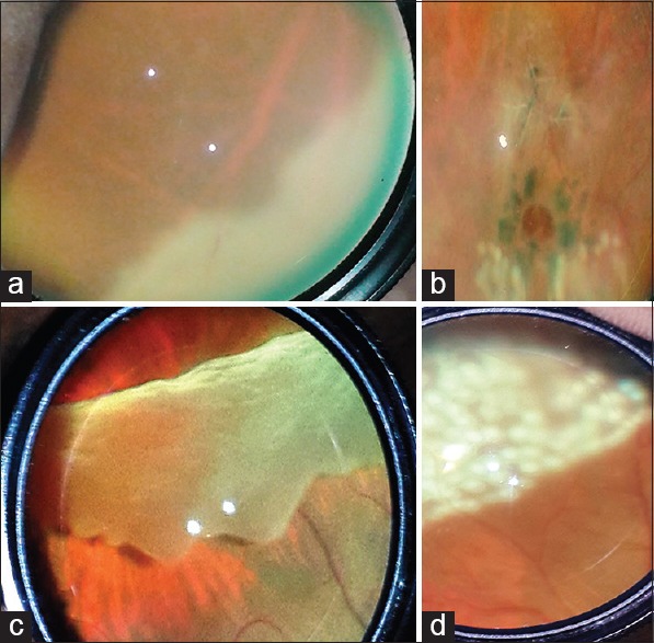
Figure 3.

Documenting peripheral retinal changes using mobile phone indirect ophthalmoscopic technique: (a) Acute retinal necrosis. (b) Lattice and atrophic hole with surrounding laser barrage. (c) Superior giant retinal tear with inversion. (d) Same eye on 1st postoperative day
