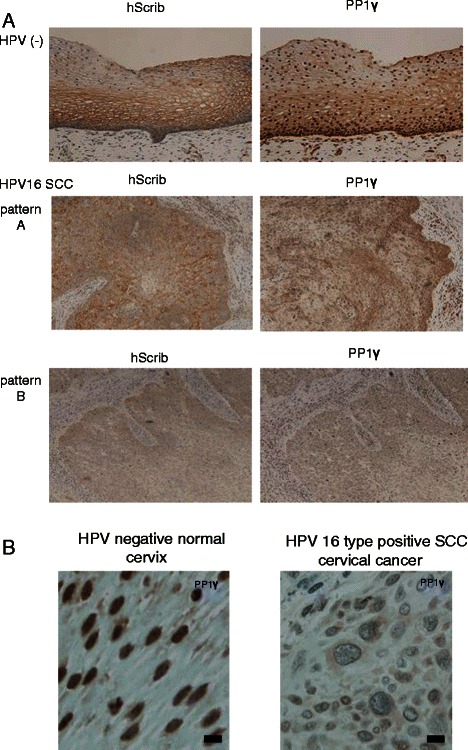
Figure 1.

Immunohistochemical analysis of the expression and localisation of hScrib and PP1γ in advanced squamous cervical carcinomas. (A) Paraffin embedded excised tissues were immunostained with anti-hScrib or anti-PP1γ as indicated, and counterstained with haematoxylin. For the antibodies, immunostaining was performed according to standard techniques using an autostainer (BenchMark XT; Ventana Medical Systems, Inc., Tucson, AZ, USA). Representative experiments for a section of cervical epitheliums from normal cervix and advanced squamous cervical carcinomas (×200 original magnification). (B) High resolution microscopic images (scale bars: 20 μm) for a section of cervical epitheliums from normal cervix and HPV-16 positive advanced squamous cervical carcinomas.
