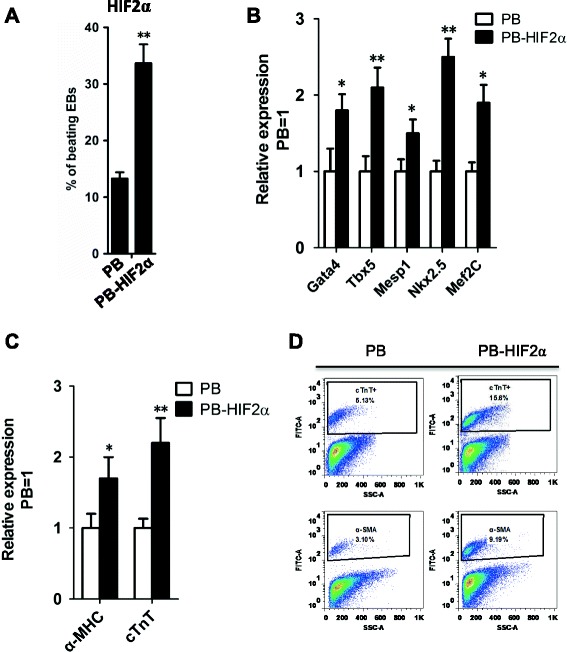
Figure 3.

HIF2α facilitated cardiac differentiation of ESCs. (A) The percentages of beating EBs derived from PB and PB-HIF2α mouse ESCs. Data represent the mean ± s.d. of three biological replicates. **p < 0.01 vs PB. (B) qRT-PCR analysis of Gata4, Tbx3, Mesp1, Nkx2.5 and Mef2c in PB and PB-HIF2α EBs at day 9. Data represent the mean ± s.d. of three biological replicates. *p < 0.05, **p < 0.01 vs PB. (C) qRT-PCR analysis of α-MHC and cTnT in PB- and PB-HIF2α EBs at day 15. Data represent the mean ± s.d. of three biological replicates. *p < 0.05, **p < 0.01 vs PB. (D) The differentiated cardiomyocytes from PB- and PB-HIF2α mouse ESCs were analyzed by FACS with cTnT and α-SMA staining.
