Figure 1.
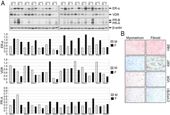
An association between higher levels of ER-α, PR-A, and PR-B and reduced levels of VDR in human UFs. A, Protein lysates were prepared from UF (F; n = 14) and adjacent myometrium (M). Equal amounts of each cell lysate (30 μg) were analyzed by Western blot for expression of ER-α, PR-A, PR-B, and VDR (top panel). Specific protein bands were quantified and normalized to β-actin, and relative values were used to generate data graphs (bottom panels). Underlining indicates UF showing up-regulation of ER-α, PR-A, and PR-B and down-regulation of VDR as compared to the adjacent myometrium. VDR and β-actin data have been published previously (19). B, Immunohistochemical analyses of a representative fibroid tumor shows reduced levels of both VDR and CYP27 as compared to adjacent myometrium. Hematoxylin and eosin (H&E) staining of UF shows tumor architecture. UF expressed higher levels of cell proliferation marker Ki67 compared to adjacent myometrium.

