Figure 6.
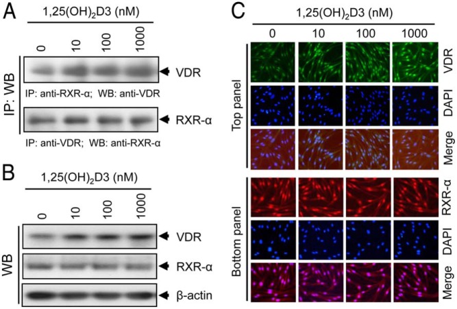
Effect of 1,25(OH)2D3 on VDR-RXR-α complex formation in cultured HuLM cells. A, HuLM cells were serum starved for 20 hours and treated with increasing doses of 1,25(OH)2D3 (0, 10, 100, and 1000 nm) for 48 hours. Cell lysates prepared from these 1,25(OH)2D3-treated cells were described in Materials and Methods. Cell lysates (1 mg) from each treatment condition were subjected to immunoprecipitation with polyclonal anti-RXR-α (top panel) or anti-VDR (bottom panel) antibodies, and immunoprecipitates were analyzed by Western blot using anti-VDR (top panel) or anti-RXR-α (bottom panel) antibodies. B, The above cell lysates were used to verify the expression of both VDR and RXR-α by Western blot analyses. C, Immunofluorescence analyses were performed using HuLM cells treated with increasing doses of 1,25(OH)2D3 for 48 hours, as described above. Cells were fixed, permeabilized, and then incubated with anti-VDR, and anti-RXR-α antibodies, followed by incubation with CY3-conjugated (red) or FITC-conjugated (green) secondary antibodies, as appropriate. Nuclei of cells were monitored by DAPI (blue). Pictures were captured at ×200 magnification.

