. 2015 Jan 16;156(4):1552–1564. doi: 10.1210/en.2014-1830

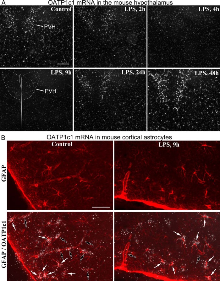
Figure 2. A, Darkfield emulsion autoradiography images demonstrate the time course of OATP1c1 mRNA expression (silver grain accumulation, white) in the mouse hypothalamus after LPS injection. Hybridization signal labeled only blood vessels but not astrocytes in the hypothalamus. The signal virtually disappears by 9 h, when only light signal remains visible in the paraventricular nucleus. At 48 h, OATP1c1 mRNA increases significantly. Scale bar = 200 μm. B, Combination of in situ hybridization for OATP1c1 (silver grains) and GFAP immunofluorescence (red) demonstrates that LPS did not affect OATP1c1 mRNA in astrocytes. Images were taken from the piriform cortex (control) and adjacent cortical amygdala (LPS). White arrows in the overlay images indicate astrocytes with OATP1c1 hybridization signal, open arrows point to blood vessel segments. Note that hybridization signal labels vessel segments in the control, but not in LPS-injected brain. Scale bar = 50 μm.

Figure 2.