. 2015 Jan 16;156(4):1552–1564. doi: 10.1210/en.2014-1830

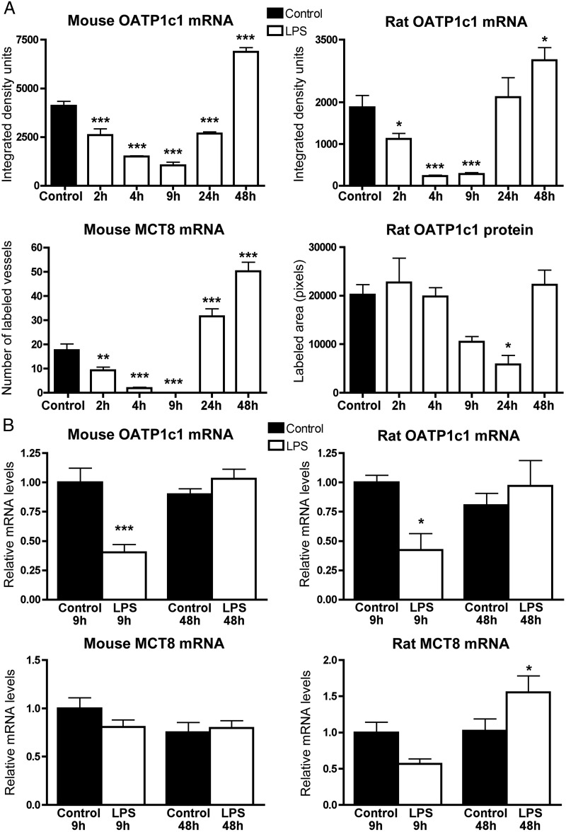
Figure 4. A, Results of semiquantitative image analysis of OATP1c1 and MCT8 in situ hybridization, and OATP1c1 immunofluorescence. OATP1c1 hybridization signal was quantified in the hypothalamus and represents mRNA expression specifically in blood vessels, as astrocytes were not labeled with the used exposure times. In the mouse MCT8 in situ hybridization experiment, the number of labeled blood vessels were counted in the diencephalic area in three sections per mouse, and the average number of vessels per section was calculated and presented in the graph. OATP1c1 immunofluorescence in rats was quantified in images taken from the thalamus by measuring the area (pixel number) covered by immunolabeled vessels. Sample sizes: four or five rats or mice/group. *, P < .05; **, P < .01; ***, P < .001 vs control. B, qPCR results from the mouse and rat cortex 9 and 48 h after LPS injection. OATP1c1 mRNA is significantly reduced 9 h after LPS injection in both species: In rats, MCT8 mRNA tended to decrease at 9 h but did not reach significance (P = .07). Sample sizes: n = 6 rats/group and n = 8 mice/group. *, P < .05, ***, P < .001 vs the corresponding controls.

Figure 4.