. 2015 Jan 16;156(4):1552–1564. doi: 10.1210/en.2014-1830

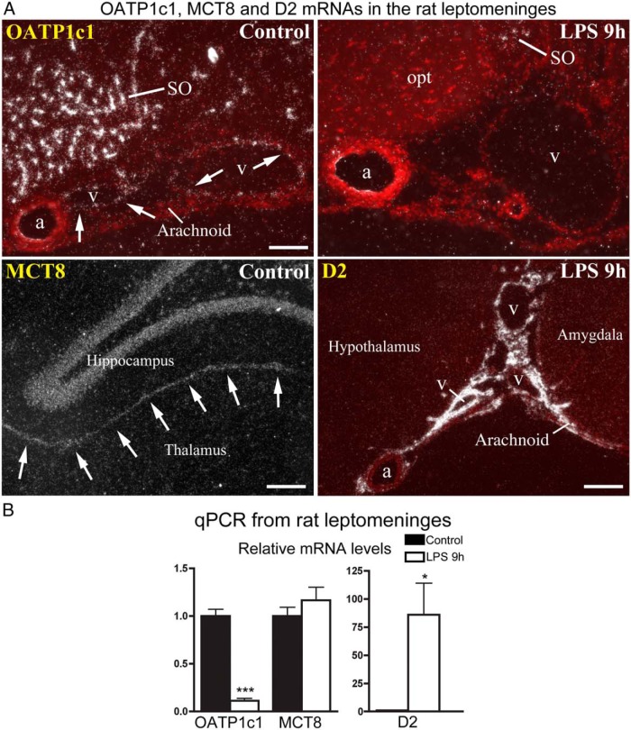
Figure 7. A, In situ hybridization for OATP1c1, MCT8, and D2 in the rat leptomeninges. Red fluorescent cresyl violet counterstaining is overlaid on darkfield emulsion autoradiography images to help identify tissue locations (OATP1c1 and D2 images). Top: OATP1c1 mRNA is expressed specifically in the wall of veins (v) that run in the arachnoid, but not in arteries (a), or the arachnoid tissue itself. Labeling of veins is absent 9 h after LPS injection. Bottom left: MCT8 mRNA is expressed in the leptomeningeal layers between the hippocampus and thalamus (arrows). Bottom right: D2 mRNA is expressed in both the arachnoid tissue and wall of arachnoid veins (v), but not arteries (a) 9 h after LPS injection. a, artery; opt, optic tract; SO, supraoptic nucleus; v, vein. Scale bar = 100 μm in top, 200 μm in bottom panels. B, qPCR results from rat leptomeningeal samples obtained from six control and eight LPS-treated rats. OATP1c1 mRNA decreases to 10% of control value, whereas D2 increases ∼86-fold on average. *, P < .05; ***, P < .0001 vs control.

Figure 7.