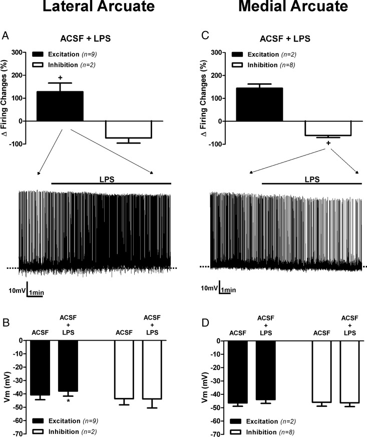
Figure 2. Contrasting effects of TLR4 activation on laterally and medially located ARC neurons.
Mean changes in firing activity (A) and mean changes membrane potential in neurons (B) located in lateral aspects of the ARC after LPS (10 μg/mL) application are shown. Mean changes in firing activity (C) and mean changes in membrane potential in neurons (D) located in medial aspects of the ARC after LPS (10 μg/mL) application are also shown. Representative examples of the most predominant response in each group is shown in the traces in panels A and C. Note that LPS increased and decreased the degree of firing activity in most neurons located in the lateral and medial ARC, respectively. +, P < .05 (paired t test within its own group). Data are represented as mean ± SEM. *, P < .05 compared with ACSF group (paired t test).

