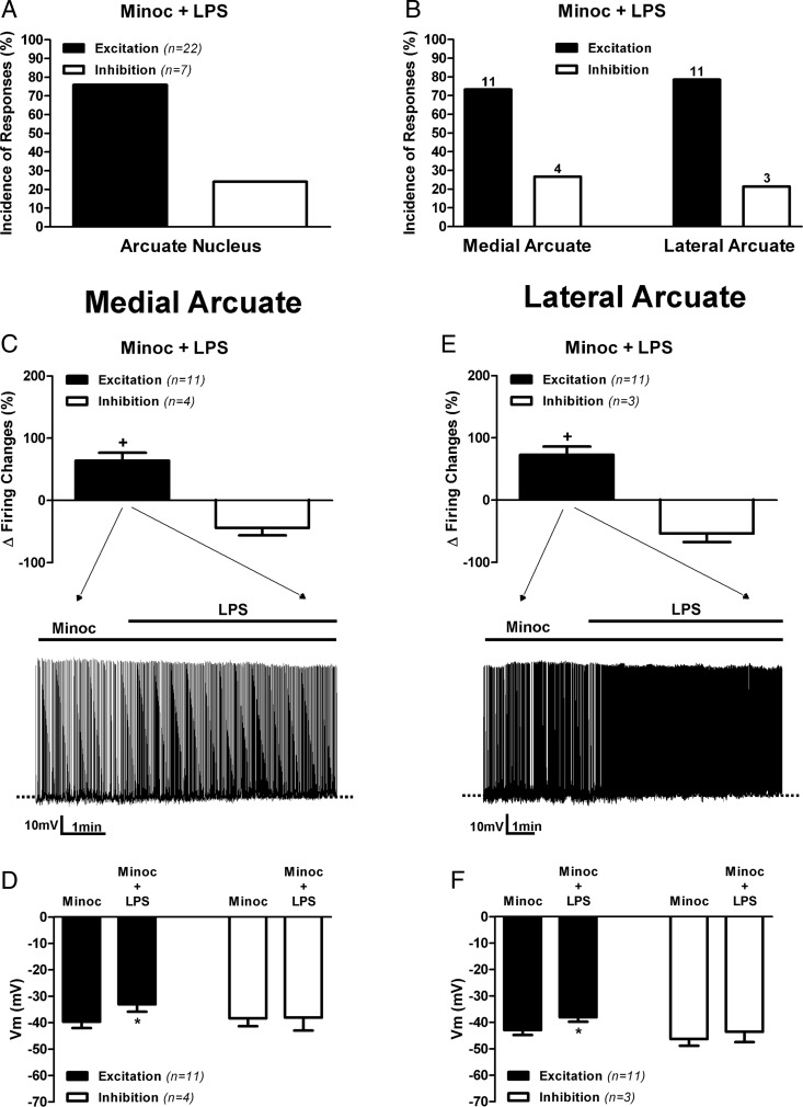
Figure 4. Inhibition of activated microglia cells prevented TLR4-mediated inhibition of medial, but no lateral, ARC neuronal activity.
Summary of the incidence of excitatory and inhibitory neuronal responses to LPS (10 μg/mL) in slices preincubated with Minoc (100 μM) observed in pooled ARC neurons (A) or according to neuronal distribution in lateral or medial ARC aspects (B). Numbers on top of bars are the number of neurons per group. Mean changes in firing activity (C) and mean changes membrane potential (D) in neurons located in medial aspects of the ARC after LPS application (10 μg/mL) in slices preincubated with Minoc (100 μM). Mean changes in firing activity (E) and mean changes in membrane potential (F) in neurons located in lateral aspects of the ARC after LPS application (10 μg/mL) in slices preincubated with Minoc (100 μM). Representative examples of the most predominant response in each group are shown in the traces in panels C and E. Data are represented as mean ± SEM. +, P < .05 (paired t test within its own group); *, P < .05 compared with Minoc (no LPS) group (paired t test).

