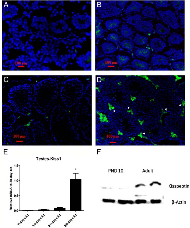
Figure 2. Developmental expression of kisspeptin in interstitial cells of mouse testes.
Histologic sections from testes of 7-day-old (A), 14-day-old (B), 21-day-old (C), and 28-day-old (D) mice were immunostained with kisspeptin antibody. Arrowheads in the photo denote KP-immunoreactive site in the tissue. E, The bar graph represents the relative mRNA expression of Kiss1 at different ages relative to 28-day-old mice (n = 4 in each group). F, Representative Western blot analysis of kisspeptin in testes lysate of 10-day-old and adult mice.

