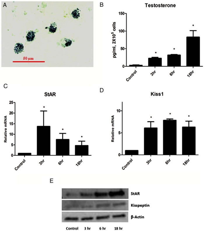
Figure 3. In vitro LH stimulation of Leydig cells.
A, 3β-HSD staining of Leydig cells isolated from the mice testes. B, Testosterone production by Leydig cells at different times after LH (20 ng/mL) stimulation. StAR (C) and Kiss1 (D) mRNA expression normalized to 18S after LH stimulation.*, significant difference compared with control. E, Western blot analysis of kisspeptin and StAR after stimulation of primary Leydig cells with LH (20 ng/mL).

