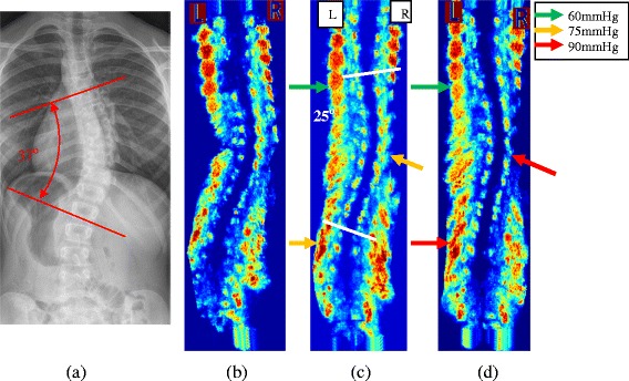
Figure 2.

X-ray and US Images of a brace patient. a) The standing pre-brace x-ray with Cobb angle 37°, b) the baseline US scan, c) the 1st trial US scan with Cobb angle 25° and d) the 2nd trial US scan with Cobb angle 23. The color arrows indicate the pressure magnitude applied to the body.
