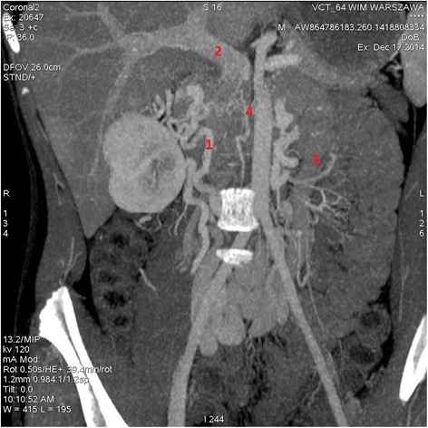
Figure 1.

CT-angiograph of 14-year old boy with absence of inferior vena cava. Scan shows lack of contrast filling at the site of vena cava inferior. Numerous veins of collateral circulation within pelvis are varicosely dilated. Because of their atypical anatomy drainage of the renal veins could not be identified.1. Renal confluence, 2. Dilated portal vein, 3. Inferior mesenteric vein, 4. Lack of contrast filling at the site of inferior vena cava.
