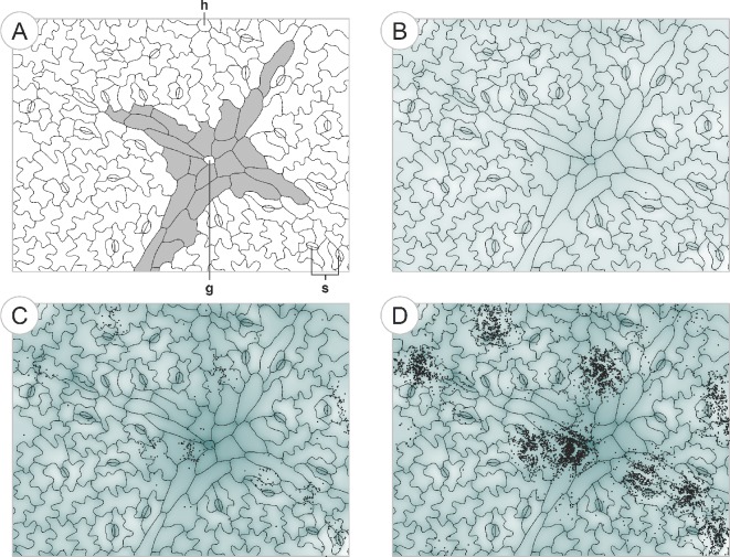
Figure 7.
A conceptual model of bacterial development based on the findings of this study. We used a simple simulation model based on the strength and range of bacterial interactions on bean leaves determined in our study in order to determine if these processes were sufficient to generate colonization patterns similar to the observed ones. The simulated colonization pattern in (D) suggests that the concert of many different interactions at different spatial scales is able to explain the complex bacterial colonization patterns observed on bean leaves. (A): the starting ‘landscape’. Epidermal structure of a young bean leaf. An x-shaped leaf vein (shaded in grey) locally approaches the leaf surface from the leaf interior with a glandular trichome (g) at the intersection. The vein is surrounded by stomata (s) and undifferentiated epidermal cells (‘puzzle pieces’). The base of a hooked trichome (h) can be seen near the middle of the upper boundary. (B): prior to colonization by microbes, resources (shaded areas) gathered on the leaf surface by processes such as epidermal leaching, especially near veins grooves between epidermal cells, and excretion by glandular trichomes. Single bacterial colonizers arrive on the leaf. (C): in the course of time, bacterial colonizers reproduce more successfully at locations rich in resources. Additionally, bacterial cells tend to get trapped near the grooves either by gravitational processes or by the increased density of leaf surface area that decreases cell motility. (D): further bacterial growth. After 20 h, growth stops in locations where the resource requirements reach the level of leaching of new resources from the leaf interior. Colonization of new resource-rich regions allows further growth of the bacterial population.

