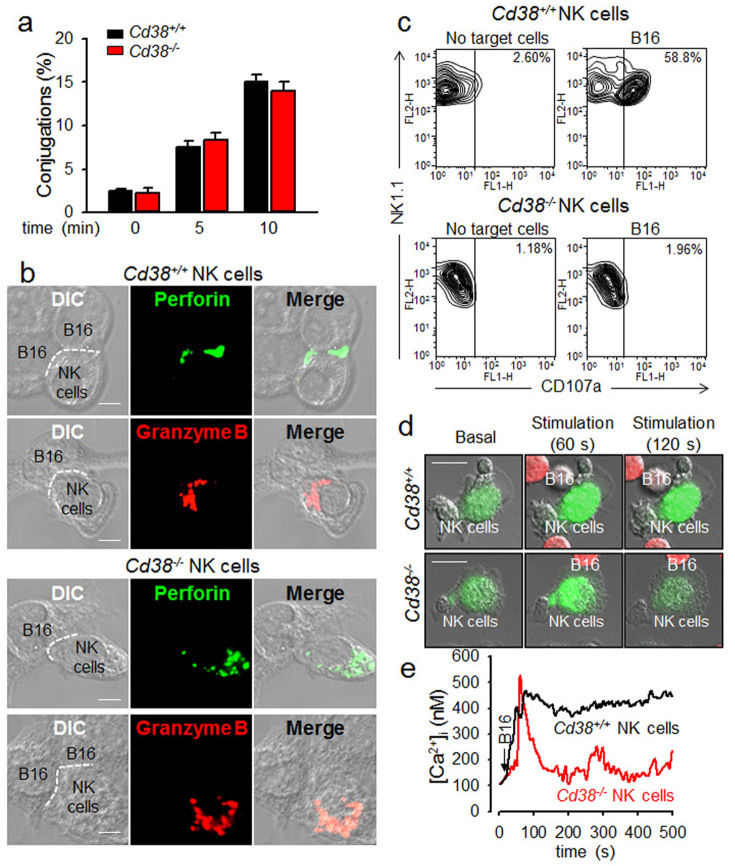
Figure 2. Tumor cell-induced NK cell granule polarization and degranulation requires CD38-mediated Ca2+ signals.
(a) Cd38+/+ or Cd38−/− NK cells stimulated with B16F10 tumor cells. The percentage of conjugation formation at indicated time was analyzed by flow cytometry. Data are mean ± SEM of three independent experiments. (b) Cd38−/− NK cells were inhibited tumor-triggered translocation of perforin and granzyme B towards immunological synapses. Cd38+/+ or Cd38−/− NK cells were stimulated with B16F10 cells at 1:1 ratio at 37°C for 20 min, and then stained with perforin or granzyme B. The dashed line indicates the immunological synapse. (Scale bar, 5 μm). All images were representative of at least three independent experiments. (c) Degranulation of Cd38+/+ or Cd38−/− NK cells upon stimulation with B16F10 cells for 2 h at 37°C. Shown is a representative of three independent experiments. (d and e) Ca2+ signals in Cd38+/+ or Cd38−/− NK cells upon stimulation with B16F10 cells. NK cells were loaded with Fluo-4 AM for 40 min at 37°C and Ca2+ levels in NK cells were measured following treatment with B16F10 cells labeled with cell tracker orange CMRA (red). Arrow indicates the time of addition of B16F10 cells. (Scale bars, 10 μm). Shown is a representative of three independent experiments.

