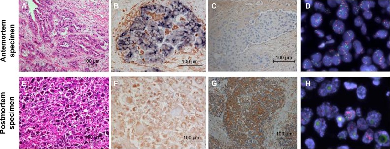
Figure 3.
Antemortem and postmortem specimens analysis.
Notes: Antemortem (A–D) and postmortem specimens (E–H) analysis. Hematoxylin and eosin staining (A and B). Double immunohistochemical staining of CDH1 (in blue) and VIM (in brown) (B and F). Immunohistochemical staining of HGF (C and G). MET gene translocation (fluorescence in situ hybridization, red signal: MET gene probe, green signal: Centromere enumeration probe 7) (D and H).
Abbreviations: CDH1, E-cadherin; HGF, hepatocyte growth factor; VIM, vimentin.

