. 2009 Jun;17(3):248–253. doi: 10.1590/S1678-77572009000300023

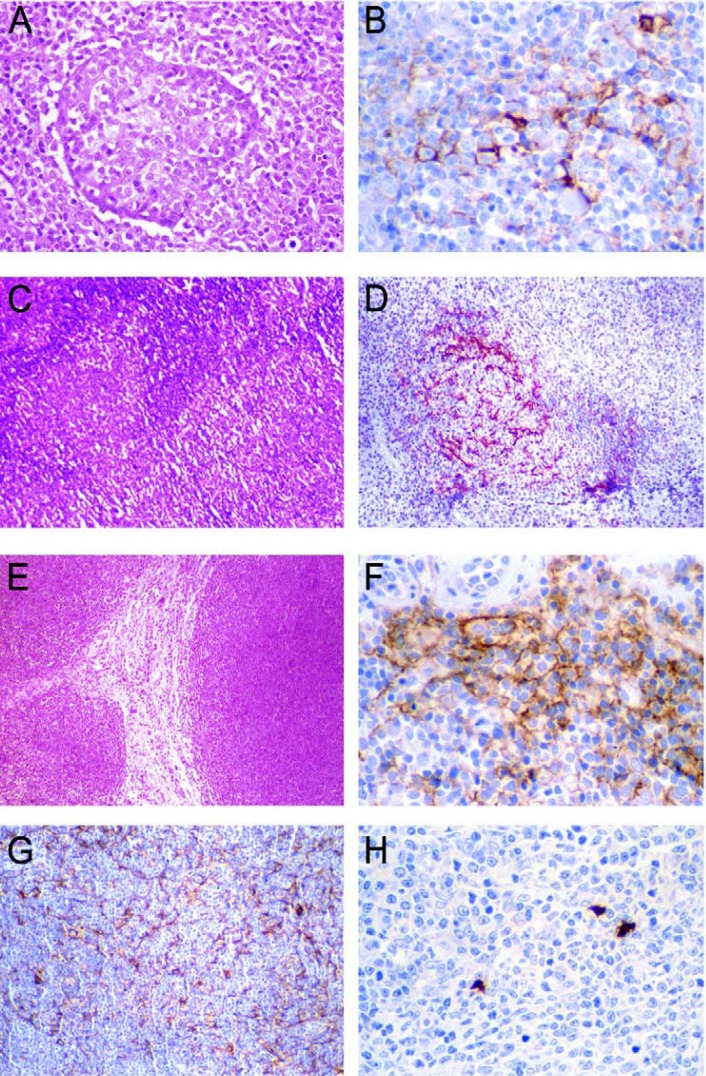
FIGURE 1. A- Pseudofollicular proliferation center in the diffuse large B-cell lymphoma (Hematoxylin-eosin, original magnification x320). B- Follicular dendritic cells stained by CD21 presented as sparse, disrupted meshwork between lymphoid cells of the pseudofollicular proliferation center of the diffuse large B-cell lymphoma (streptavidin-biotin, original magnification x640). C- MALT lymphoma presenting reactive follicles (Hematoxylin-eosin, original magnification x100). D- Colonized follicles by follicular dendritic cells. A meshwork is revealed by CD21 in the MALT lymphoma (streptavidin-biotin, original magnification x320). E- Mantle cell lymphoma demonstrates a monomorphic lymphoid proliferation with a nodular pattern (Hematoxylin-eosin, original magnification x100). F- CD21 demonstrates loosely arranged, ill-defined and expanded meshwork of follicular dendritic cells at the periphery of the mantle cell lymphoma (streptavidin-biotin, original magnification x640). G- Caldesmon revealed a dense and confluent meshwork of the follicular dendritic cells among lymphoid cells of in a case of diffuse large B-cell lymphoma (streptavidin-biotin, original magnification x200). H- Interdigitating dendritic cells revealed by S100 protein in one case of the diffuse large B-cell lymphoma are visualized as irregular spots (streptavidin-biotin, original magnification x320).

FIGURE 1