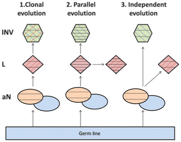
Figure 5.

Model of lineage between adjacent lepidic and invasive adenocarcinomacomponents.Demonstrates the three models of lineage between adjacent tissues. 1, the clonal evolution model describes a direct progression from the in situ lepidic tumor components (red) to the invasive tumors (green). 2, the parallel evolution model introduces the further concept that the transition took place some time in the past and both the lepidic and invasive components are continuing to progress and accumulate further unique mutations. 3, the independent evolution model predicts the lepidic and invasive components to grow independently from a common background. Each model also incorporates the concept of the associated normal (aN) in the local environment from where the tumors emerged. This aN is split into two colors to indicate the germline (gray blue) and the abnormal (orange), which has accumulated background mutations above the germline but would be common to any tumor arising from that environment. Each component additionally contains black horizontal, red vertical, and/or blue vertical lines to represent mutations occurring in aN, lepidic, or invasive, respectively.
