Figure 1.

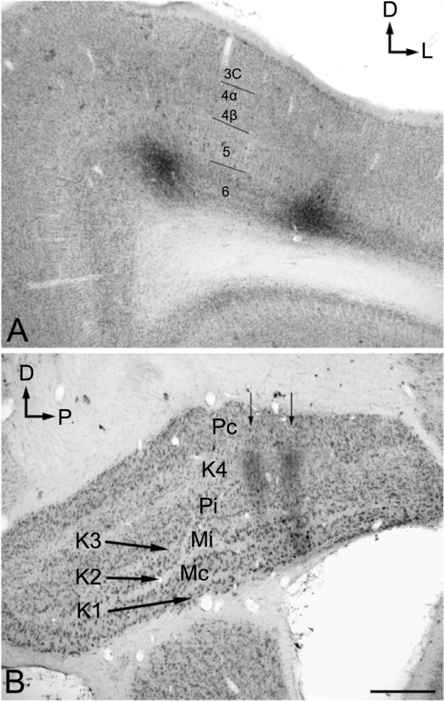
Photomicrographs of two iontophoretic BDA injection sites.
Notes: (A) Coronal section through the dorsal bank of V1. (B) The transported label in a parasagittal section through the LGN from the same case. The injections were centered in cortical layer 6 and confined to the infragranular layers of V1 (eg, layer 4 and 5). Here, we used cortical layer designations as follows with Brodmann’s19 terminology in parenthesis where it differs: 3C (4B), 4α (4Cα), 4β (4Cβ). The rationale for this choice is given in Casagrande and Kaas.18 The labeled axons in the LGN form two columns (arrows) that correspond to the pair of injections in layer 6 of V1. Numerals in LGN indicate different K layers. Scale bar =500 μm for (A) and (B).
Abbreviations: BDA, biotinylated dextran; D, dorsal; K, koniocellular; L, lateral; LGN, lateral geniculate nucleus; Mc, contralateral magnocellular; Mi, ipsilateral magnocellular; P, posterior; Pc, contralateral parvocellular; Pi, ipsilateral parvocellular; V1, primary visual cortex.
