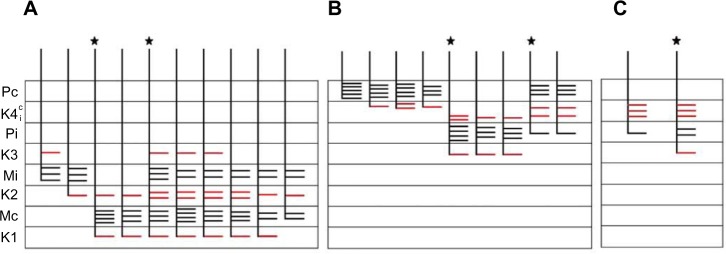
Figure 5.
Schematic diagrams summarizing the branching patterns of corticogeniculate axons.
Notes: Corticogeniculate axons reconstructed in the (A) M layers (n=10), (B) P layers (n=9), and (C) K layers (n=2). The number and length of bars are not intended to represent exact branch length, but give a general idea of branch density within each LGN layer. The axons marked with a ★ are also shown in Figure 4 (A), Figure 6 (B) and Figure 7 (C). Branches that terminate in K layers for every axon are indicated in red.
Abbreviations: K, koniocellular; , koniocellular contralateral and ipsilateral portions of layer; LGN, lateral geniculate nucleus; M, magnocellular; Mc, contralateral magnocellular; Mi, ipsilateral magnocellular; P, parvocellular; Pc, contralateral parvocellular; Pi, ipsilateral parvocellular.

