
FIGURE 2.
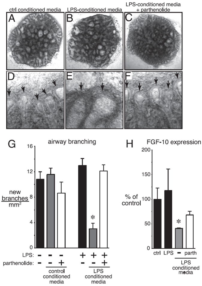
LPS-conditioned media disrupt saccular airway branching in TLR4 mutant explants. Media from control and LPS-treated BALB/cJ explants were added to LPS-resistant C.C3-Tlr4Lpsd/J explants. A–F, Brightfield images of C.C3-Tlr4Lpsd/J explants cultured with control-conditioned media (A, D), LPS-conditioned media (B, E), or LPS-conditioned media with the NF-κB inhibitor parthenolide (1 μM; C, F) (original magnification ×25). Higher-magnification images are shown in D–F (original magnification ×200). Arrows indicate saccular airways. G, LPS-conditioned media inhibited formation of new saccular airways in C.C3-Tlr4Lpsd/J explants. *p < 0.001; n = 7. H, LPS-conditioned media inhibited FGF-10 expression in C.C3-Tlr4Lpsd/J explants, as measured by real-time PCR. *p < 0.05; n = 9.