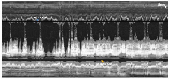
Fig. 6.

En face OCT image from acquired volume with the fast distal rotary scan and no longitudinal actuation in order to assess motion artifacts. Large hyposcattering (dark) regions indicated areas where the esophageal wall is not in contact with the capsule. The horizontal axis corresponds to time and spans 28 seconds. Periodic motion artifacts are consistent with cardiac and respiratory cycles.
