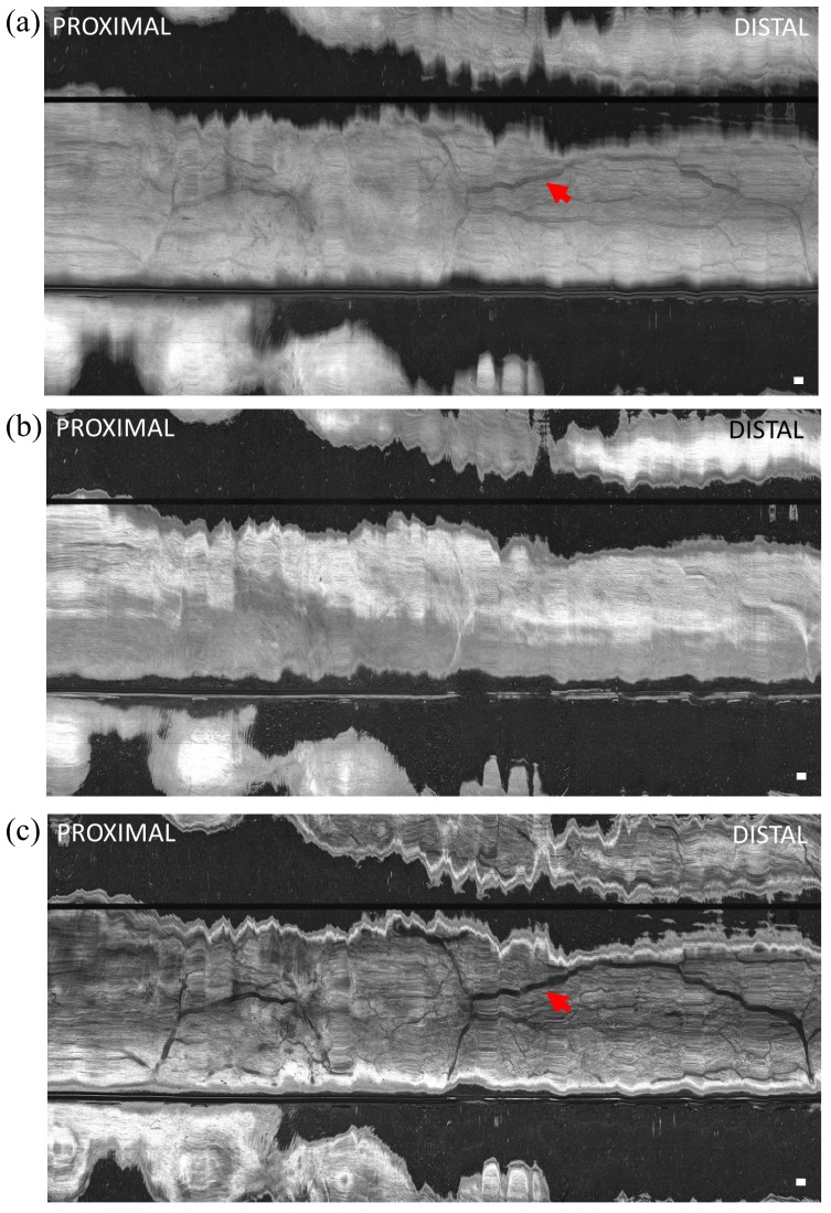
Fig. 7.
En face OCT image of swine esophagus in vivo (a) projected over 600 μm depth range from the tissue surface, (b) at 200 μm below the surface and projected over 100 μm depth range, and (c) at 600 μm below the surface and projected over 100 μm depth range. The capsule was manually advanced for the longitudinal scan. Shadow patterns resembling a vascular network (red arrows) were visible. The horizontal scale was based on an estimate of manual longitudinal pullback speed of ~3 mm/s. Scale bar is 1 mm.

