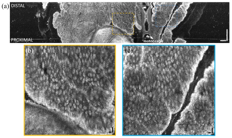
Fig. 9.
(a) En face OCT image of swine rectum at ~400 μm depth from tissue surface and projected over ~50 μm depth range. Insets (b, c) show enlargements of regions demonstrating the high precision distal pneumatic scanning. Non-uniform rotational distortion (NURD) of the micromotor was visible. Scale bars of large image and insets are 1 mm and 100 µm, respectively. Scanned area was approximately 1.3 cm2. The motor cable (covered by plastic extrusion on proximal end cap) appeared aliased at a different depth as indicated.

