
Fig. 7.
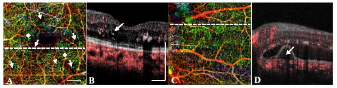
(A) wOMAG of the left eye in the DR patient showing pathologies including capillary drop-out (arrows), tortuosity (arrowhead) and possible microaneurysm (diamond). (B) Structural OCT scanned at the dotted line of (A) showing the coexistence of an edema in the region with significant capillary dropout. (C) wOMAG of the left eye in the PCV patient. (D) Structural OCT scanned at the dotted line of (C) showing the existence of a pigment epithelial detachment below the hyper-reflective foci. The scale bar in (A) and (B) indicates 500 μm.