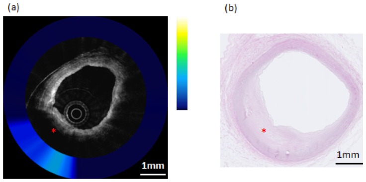
Fig. 7.

Composite OCT-NIRAF image and corresponding histology for a non-necrotic lipid containing plaque (Pathological Intimal Thickening–PIT). a) The OCT image indicates the presence of an area of high attenuation suggestive of lipid pool (star). The NIRAF signal is elevated, but only moderately so, over the lipid pool location. b) H&E stained slide confirming that this lesion (star) is pathological intimal thickening. The scale bars for both (a) and (b) represent 1mm.
