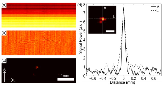
Fig. 2.

Results from a 7 µm carbon fiber at 4.5 mm depth. (a) Raw image. (b) Image after cross-talk removed. (c) Image after cross-talk removed and reconstruction, with axial (A) and lateral (L) dimensions labelled. (d) Axial and lateral cross-sections taken through the reconstructed image shown in (c) with profile inset (scale bar = 200 µm).
