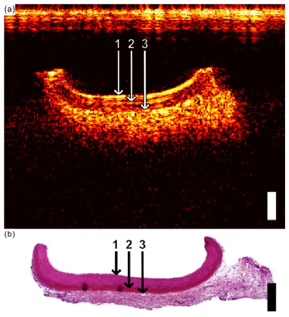
Fig. 5.

(a) Optical ultrasound image of the carotid artery, showing the tunica media (1 = top boundary; 2 = bottom boundary), the external elastic lamina (2 = top boundary; 3 = bottom boundary), and the adventitia (3 = top boundary). (b) Corresponding histological cross-section (H&E) of the carotid artery from which the layers in (a) were interpreted. In both (a), and (b), the scale bar corresponds to 1 mm for both axial and lateral dimensions.
