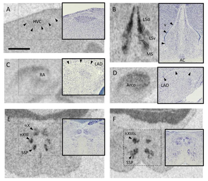
Fig. 2.
Examples of oxytocin-like receptor (OTR) mRNA signal quantified in this study. Each panel shows a film autoradiogram along with an inset depicting a Nissl-stained alternate section from the same animal. All photos are from a testosterone-treated male. Scale bar in A, 1mm, applies to all images. A. HVC (used as a proper name). Midline is to the right. B. The lateral septum, showing the dorsal (LSd) and ventral (LSv) subdivisions, and the medial septum (MS) Variation in the OTR mRNA signal was used to delineate the areas, which are not distinct in Nissl-stained sections (Goodson et al., 2004). v, lateral ventricle. C. The robust nucleus of the arcopallium (RA) was relatively unlabeled compared with the surrounding arcopallium. LAD, dorsal arcopallial lamina. Midline to the right. D. The arcopallium was examined in sections caudal to RA. Midline to the right. E. The lingual portion of the hypoglossal nucleus (nXIIl) can be seen ventral to the motor nucleus of the vagus (nX) and dorsal to the supraspinal nucleus (SSP). F. The tracheosyringeal portion of the hypoglossal nucleus (nXIIts) is positive for OTR mRNA.

