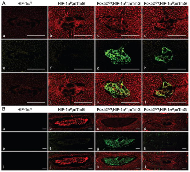
Figure 2.
Lineage study in control and mutant IVDs. A, B. Detection of fluorescence in frozen sections of NP isolated from E15.5 (A) and 1 month (B) HIF-1αf/f (a,e,i), HIF-1αf/f;mTmG (b,f,j), Foxa2iCre;HIF-1αf/+;mTmG (c,g,k) and Foxa2iCre;HIF-1αf/f;mTmG (d,h,l) mice, respectively. Red fluorescence (a–d), green fluorescence (e–h) and merged filters (i–l) are shown. Red signal is membrane-bound tdtomato fluorescent protein indicating cells in which recombination has not occurred. Green signal is membrane-bound EGFP indicating cells in which Cre mediated recombination has occurred. Bar = 100 μm. Image reproduced with permission from Merceron C, et al. 2014. Loss of HIF-1α in the Notochord Results in Cell Death and Complete Disappearance of the Nucleus Pulposus. PLoS One. 9:e110768.

