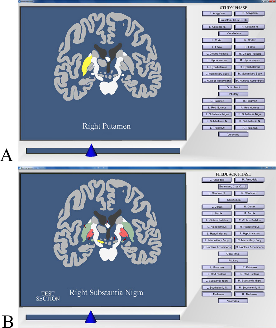
Figure 2.
Screen images from the program for learning sectional anatomy. A: Image from the study phase of a trial. The learner has moved to a coronal section that cuts through the middle of the brain and has highlighted the right putamen. B: Image showing post-test graphical feedback. Tested items are indicated by small red arrows. Structures colored green were labeled correctly on the test, structures colored red were mislabeled, and achromatic structures indicated by the red arrows were skipped. The learner has highlighted the right substantia nigra.

