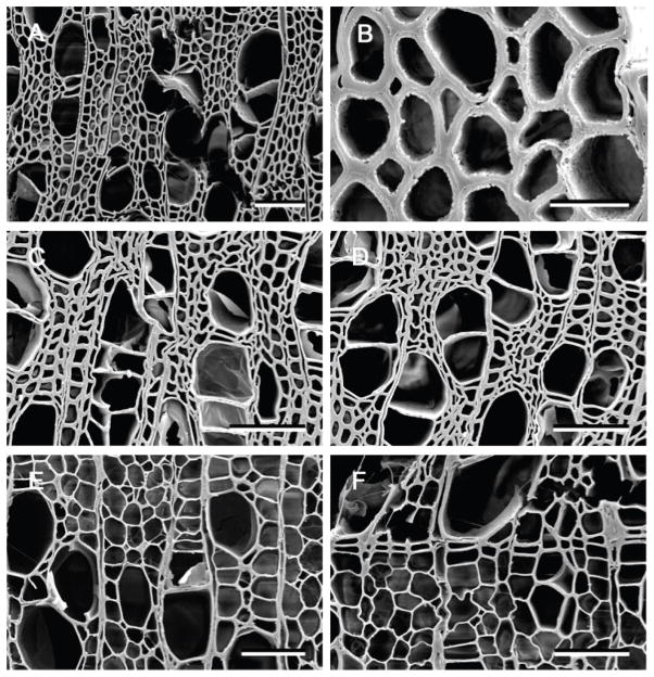
Figure 5.
Scanning electron micrographs of transverse sections of aspen (Populus) wood decay by Cylindrobasidium torrendii (A and B), Fistulina hepatica (C and D) and Schizophyllum commune (E and F). A and B. Localized degradation of all cell wall components with erosion of the wall taking place from the cell lumen towards the middle lamella. Small voids occurred in the wood cells where all cell wall layers were degraded. C and D. A diffuse attack on wood cells resulted in cells with altered walls. No cell wall erosion took place but walls were slightly swollen and cells were partially collapsed and appeared convoluted. E and F. Thinning and eroded secondary wall layers were evident in wood cells. In some cells, the secondary wall was completely degraded but the middle lamella between cells remained. The thinned cell wall broke and detached in some areas resulting in small voids. Bar = 100 μm in A, 20 μm in B and 50 μm in C, D, E, F.

