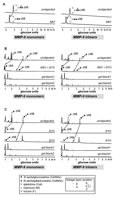
Figure 5. Glycan analysis of proMMP-9 monomers and trimers.
(A) N-linked glycans of separated proMMP-9 monomer and trimer preparations were isolated and analyzed by HILIC. Typical profiles of undigested and BKF-digested monomeric and trimeric proMMP-9 N-glycomes are shown. (B, C) Typical HILIC profiles of monomeric and trimeric proMMP-9 O-glycomes before and after digestion with a range of exoglycosidases. Peaks were collected and each digested with BTG and/or ABS. Structural symbols for the glycans and their linkages are shown in the box situated at the bottom of the figure. These symbols are also the keys to the structures shown in Table 1.

