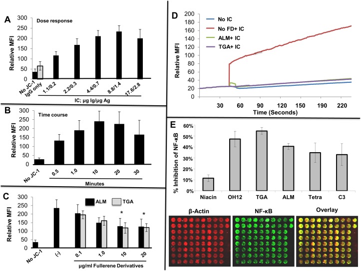
Fig 2. Mitochondrial membrane potential correlates with MC degranulation through FcγR receptors and is inhibited by fullerene derivatives.
In Fig 2A, change in mitochondrial membrane potential as a function of the concentration of IC stimulus was assessed. Human MCs were stimulated with graded concentrations of preformed IgG anti-NP/NP-BSA immune complexes as indicated for 10 minutes. As a control, cells without JC-1 and cells with JC-1 plus NP IgG only (no antigen) were incubated in parallel. The above experiment is representative of two separate samples. The percent of degranulation from these cells was 23%, 32%, 39%, and 45% respectively. Mitochondrial membrane polarization was quantified by cytofluorimetry (FL2 channel) using FACs analysis as described above. As seen in Fig 2B, change in mitochondrial membrane potential as a function of time with fixed concentration of IC stimulus was assessed. Human MCs were stimulated with 8.8 μg/ml anti-NP Ab with 0.13 μg/ml NP-BSA of preformed IgG anti-NP/NP-BSA IC for the indicated times. As a control, cells without JC-1 were incubated in parallel. In Fig 2C, Fullerene derivatives inhibit IC-induced increases in mitochondrial membrane potential. Mast cells were incubated overnight with ALM or TGA (10 μg/ml) or media only. The next day cells were challenged with media containing JC-1 probe for 10 minutes at 37°C with or without IC (as in A). After 10 minutes cells were washed with cold PBS, centrifuged and the JC-1 aggregates detected using the FL2. The above experiment is representative of three separate samples. As shown in Fig 2D fullerene derivatives inhibit IC-induced elevations in intracellular ROS levels. Mast cells were incubated overnight with fullerene derivatives, washed and DCF-DA added to cells for 30 minutes at 37°C. After washing cells were activated with optimal concentrations of IC and the fluorescence intensity measured at 525nm after establishing baseline. Figs. show representative numbers from duplicate samples for each condition and are representative of three separate MC cultures. Fig 2E shows that fullerene derivatives can block Fcγ receptor mediated activation of the MC transcription factor NF-κB. Mast cells were incubated with or without fullerene derivatives (10 μg/ml) overnight, washed, and challenged with IC for 24 hours. After washing, in-cell Westerns were performed using the manufacturers protocol. Control wells (those without primary antibodies) were reserved as a source for background well intensity. Further controls were cells incubated without fullerene derivatives or IC. Results represent results from two separate experiments.

