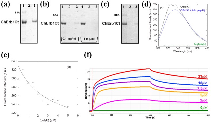
Fig 8. ChErb1Ct (residues 432–801) binds RNA in vitro.
(a) Coomassie-stained SDS-PAGE showing the binding of Erb1 β-propeller from Ch. thermophilum to polyU agarose beads. (b) The saturation of the binding is visible upon addition of 0.1 mg/ml or 1 mg/ml of free polyuridilic acid. (c) The binding is still detectable upon addition of 1 mg/ml of heparin to the binding buffer. (a) (b) and (c) 1: Input, 2: Wash, 3: Elution; (d) Fluorescence spectra of ChErb1432-801 alone (in black) and with 5μM poly(U) (blue) obtained by excitation at 280 nm. The spectra were acquired at 25°C with 1.5 μM of ChErb1432-801. The green line at the bottom of the spectra is the emission spectra of polyU at a concentration of 5 μM. (e) Titration curve obtained from the emission fluorescence intensity at 315 nm. The line is the fitting to Eq (1). (f) Association and dissociation curves of 15nt-poly(U) with different concentrations of ChErb1432-801 measured by biolayer interferometry.

