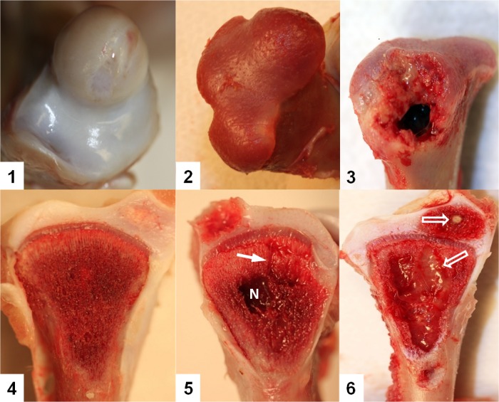
Fig 1. Stages of proximal femoral head degeneration (Upper row) or tibial head necrosis (Low row) leading progressively to bacterial chondronecrosis with osteomyelitis (BCO).
1. Normal proximal femoral head; 2. Femoral head separation (FHS: epiphyseolysis); 3. Femoral head necrosis, FHN. 4. Normal proximal tibial head with struts of trabecular bone in the metaphyseal zone fully supporting the growth plate; 5. Tibial head necrosis (THN). Lytic channels (small arrows) penetrate from the necrotic voids into the growth plate. 6. Tibial head necrosis (THNsc). Bacterial infiltration and sequestrae (open arrows) provide macroscopic evidence of osteomyelitis.

