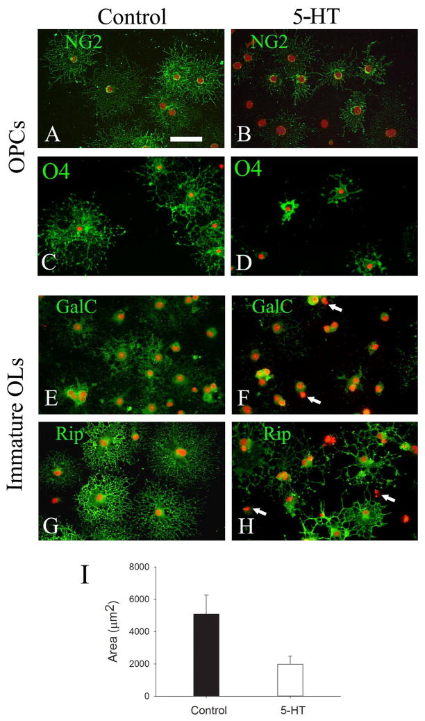
Fig. 3.
5-HT exposure led to OL developmental disturbance. OPCs (A-D) or Immature OLs (EH) were treated with 5-HT (10 μm) for 5 days, and cells were immunostained with stage-specific OL markers to assess morphological changes. OPCs treated with 5-HT showed significantly reduced NG2 and O4 immunoreactivity (B and D, respectively), as compared to control cells (A and C, respectively). Compared to OPCs, immature OLs showed even greater developmental disturbance, indicated by not only weaker GalC immunostaining in the somata, but also markedly reduced processes in the 5-HT treatment (F), as compared to the control (E). The compromised processes in 5-HT treated cells were more pronounced by Rip immunostaining, which showed fewer, shortened, and distorted processes (H), compared to elaborating, extensive process network in the control (G). Some degenerative cells in 5-HT-treatment are shown by their condensed nuclei (marked as arrows in F and H). Semi-quantitative analysis of Rip immunolabeling showed that 5-HT treatment significant reduced cell surface areas (I). Data were from a total of 50 cells from three independent experiments. *p<0.01 vs the control. Scale bar: 50 μm.

