
Fig. 4.
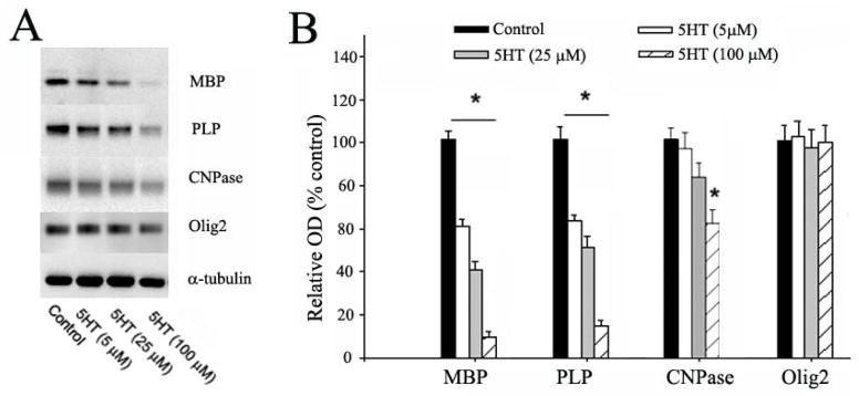
5-HT exposure reduced the expression of myelin proteins in OLs. A: representative Western blots show the expression of MBP, PLP, CNPase, and Olig2 in immature OLs following exposure to 5-HT (5–100 μM) for 5 days. B: The relative expression levels of myelin proteins were determined by normalizing the optical density (OD) of their respective target bands to α-tubulin, expressed as a percentage to the control. The expression of MBP and PLP was significantly reduced by 5-HT exposure in a dose-dependent manner. In contrast, a significant reduction of CNPase expression was only found when 5-HT concentration reached 100 μM, whereas no change of Olig2 expression was detected at all doses. *p<0.05 vs the control. Data are mean ±SD from three independent treatments.