Fig. 5.
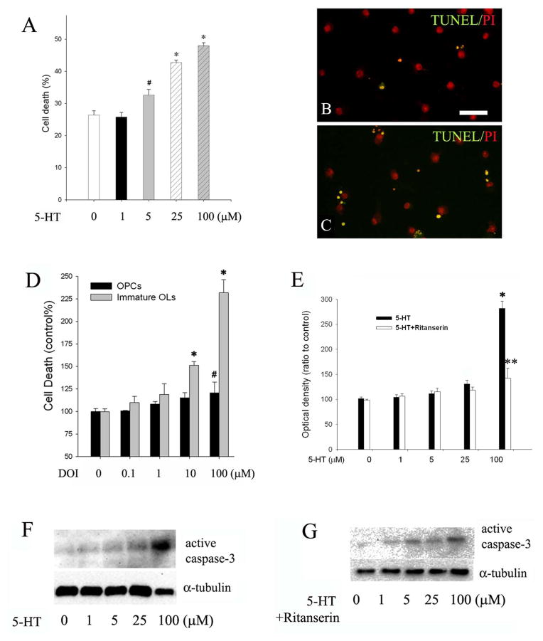
5-HT-induced OL death was mediated by 5-HT2A receptor. Immature OLs were exposed to increasing concentration of 5-HT for 48 h, and cell death was determined by TUNEL. A: the percentage of TUNEL+ cells increased significantly in 5-HT treatment, in a dose-dependent manner. B-C: representative micrographs of TUNEL (green fluorescence) in the control (B) and 5-HT treatment (C, shown 5-HT at 25 μM). Nuclei were counter-stained with PI (red). Scale bar: 50 μm. D: exposure to 5-HT2A receptor agonist DOI led to cell death in both OPCs and immature OLs; however, immature OLs showed an increased susceptibility than OPCs. *p<0.01 and #p<0.05 vs their respective controls (shown as 0 μM). E: conversely, 5-HT-induced caspase-3 activation in immature OLs was blocked by pre-treatment with ritanserin (1 μM). **p<0.01 vs 5-HT at 100 μM. F–G: representative western blots show that 5-HT dose-dependently activated caspase-3 in immature OLs (F), which was partially blocked by pre-treatment with ritanserin. Data are presented as mean ±SD from three independent experiments.

