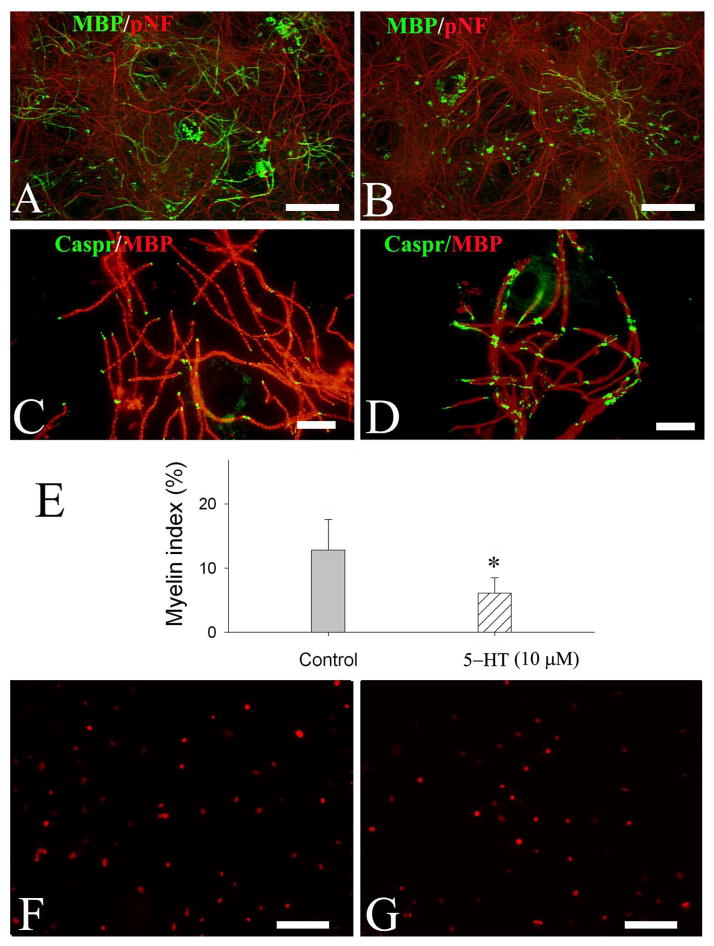
Fig. 6.
5-HT exposure resulted in myelin malformation in neuron-OL co-cultures. Neuron-OL myelination cultures were exposed to 5-HT (10 and 100 μM) from DIV10 to DIV40. Myelinated internodes were revealed by co-localization of MBP and pNF immunostaining, and the paranodal domains were demonstrated by Caspr immunofluorescence. Representative micrographs show myelinated internodes (double-labeling with MBP and pNF) in the control (A) and 5-HT-treated cultures (B). The expression of caspr in 5-HT-treated cultures showed a diffuse, enlarged pattern (D), which was in contrast to a more localized, regular spaced pattern in the control culture (C). Quantification of myelinated internodes shows that myelin index was significantly reduced by 5-HT exposure (E); however, no significant difference was noted between 10 and 100 μM 5-HT treatments. There was no significant change of total OL lineage numbers between 5-HT treatment (G) and the control (F). Data were from three independent treatments with triplicate coverslips per condition. Scale bars: 200 μm (A, B, F&G); 50 μm (C&D).

