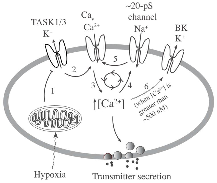
Figure 2. A model showing a critical role of TASK in hypoxia-induced secretion of transmitters.
Hypoxia acts on the O2 sensor (probably mitochondria) that generates a signal that inhibits TASK activity (step 1). Inhibition of TASK leads to cell depolarization due to continuous influx of Na+ via background Na+ channels and this opens Ca2+ channels (step 2). Ca2+ influx increases intracellular [Ca2+] in the cell (step 3) stimulating the secretion of transmitters. Ca2+ acts on the 20-pS cation channel (NSCC) to increase Na+ influx (step 4) and enhance Ca2+ influx (step 5). High [Ca2+]i activates BK to limit further depolarization (step 6). These mechanisms maintain the level of depolarization and [Ca2+]i in response to a given level of hypoxia.

