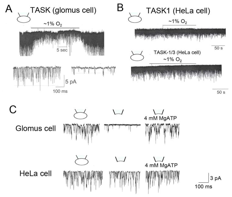
Figure 4. O2 and ATP sensitivity of TASK expressed in glomus cells and HeLa cells.
(A) Tracing shows a rapid and reversible inhibition of TASK activity in response to hypoxia in glomus cells. Channel currents are recorded from cell-attached patches with pipette potential at 0 mV. Pipette solution contains (mM): 140 KCl, 5 EGTA, 1 MgCl2 and 10 HEPES (pH 7.3) and the bath perfusion solution contains (mM): 117 NaCl, 5 KCl, 23 NaHCO3, 1 MgCl2, 1 CaCl2, 10 HEPES and 11 glucose (pH 7.3). Expanded current tracings show that hypoxia reduces TASK activity as well as the amplitude. (B) Same experiment as in A except that HeLa cells expressing TASK1 or TASK1/3 are used for channel current recording. Hypoxia has no effect on TASK activity. (C) Cell-attached patch recording from glomus cells shows robust TASK activity. TASK activity decreases markedly upon formation of inside-patch in glomus cells but not in HEK293 cells. Application of MgATP restores TASK activity in glomus cells.

