Fig. 1.

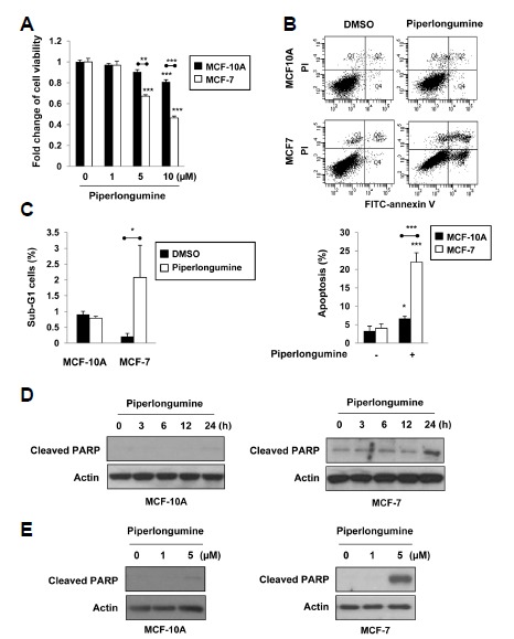
Piperlongumine induces apoptosis in human breast cancer MCF-7 cells relative to human MCF-10A breast epithelial cells. (A) MCF-10A and MCF-7 cells were treated with 0, 1, 5 or 10 μM of piperlongumine for 24 h. Cell viability was evaluated by the MTT assay. (B, C) Cells were treated with 5 μM of piperlongumine for 36 h. Quantification of apoptosis was determined by flow cytometry (B). The proportion of the cells at the sub-G1 phase was also assessed by flow cytometry analysis as described in the Materials and Methods section (C). Means ± S.D. (n=3), *p < 0.05, **p < 0.01, ***p < 0.001. (D, E) MCF-10A and MCF-7 cells were treated with 5 μM of piperlongumine for the indicated time periods (D) and 0, 1 or 5 μM of piperlongumine for 24 h (E). Total protein isolated from cell lysates was subjected to immunoblot analysis for the measurement of cleaved PARP. Actin was used as an equal loading control for normalization.
