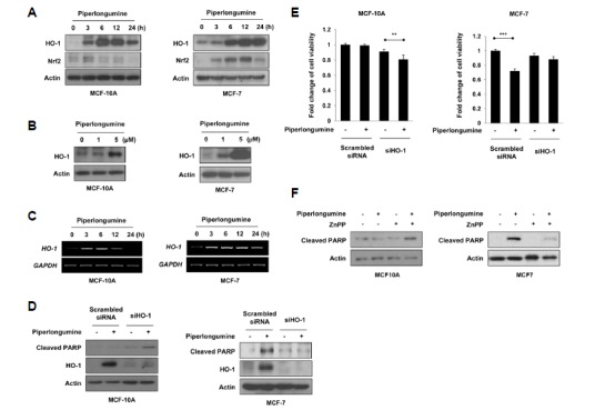
Fig. 2.

HO-1 mediates the selective effect of piperlongumine on cancer cell apoptosis. (A-C) MCF-10A and MCF-7 cells were treated with 5 μM of piperlongumine for the indicated time periods (A, C) and 0, 1 or 5 μM of piperlongumine for 24 h (B). (A, B) Total protein isolated from cell lysates was subjected to immunoblot analysis for the measurement of HO-1 and Nrf2. Actin was used as an equal loading control for normalization. (C) The expression of HO-1 mRNA was determined by semi-quantitative RT-PCR. The level of GAPDH mRNA was used as an internal control. (D, E) Cells were transfected with scrambled or HO-1 siRNA for 24 h, and then exposed to piperlongumine for additional 24 h. The protein levels of cleaved PAPR, HO-1 and actin were determined by Western blot analysis (D). Cell viability was evaluated by the MTT assay (E). Means ± S.D. (n = 3), **p < 0.01, ***p < 0.001. (F) MCF-10A and MCF-7 cells were treated with piperlongumine in the absence or presence of ZnPP for 24 h. The protein levels of cleaved PAPR, HO-1 and actin were determined by Western blot analysis.
