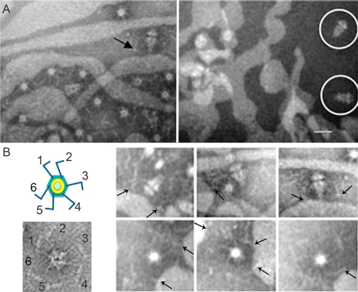
FIGURE 2.
T7 bacteriophage interacts with rough LPS trough the fiber protein. A, electron micrographs showing negatively stained fibered (left) or fiber-less tail complexes (right) incubated with LPS. The arrow indicates representative tail complex showing interaction between the fiber and rough LPS; a white circle surrounds non-fibered complexes. Bar = 30 nm. B, left, uranyl acetate negatively stained end-on view tail complex where the six protruding fibers were labeled. The scheme on the top represents a tail complex where the tubular structure has been colored in green (tail proteins) and yellow (connector), and the fibers have been colored in blue. Right, gallery showing different tail complexes in end-on and side views interacting with the LPS. Arrows point to representative fiber complexes.

