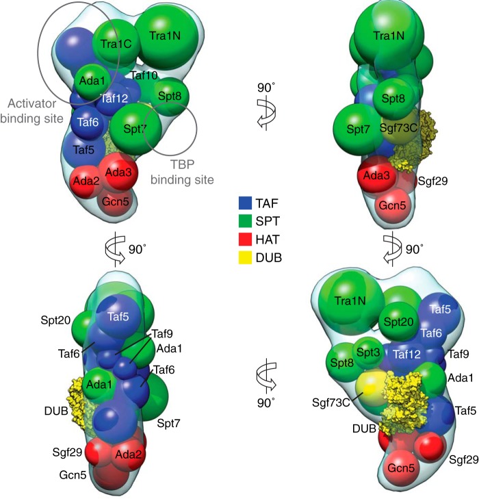
FIGURE 7.
Spatial arrangement of SAGA subunits. Results from EM-based subunit localization experiments and cross-linking mass spectrometry were combined with the curved SAGA three-dimensional reconstruction to propose an overall arrangement of all 19 complex subunits. The volumes of spheres represent the molecular mass of each subunit (DUB module crystal structure, Protein Data Bank code 3M99).

