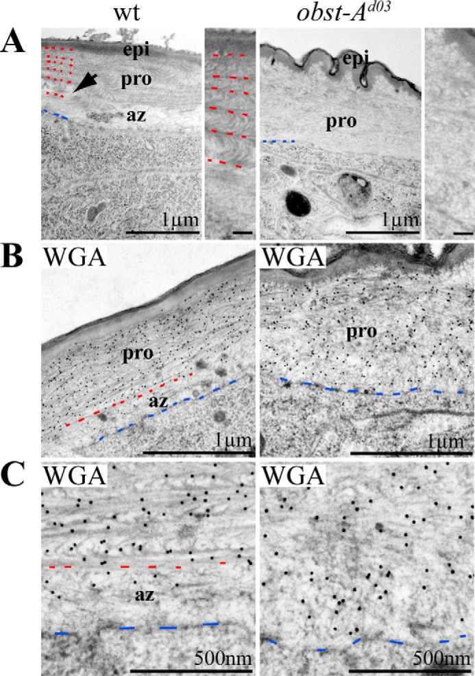
FIGURE 2.

Obst-A organizes chitin matrix structure at the assembly zone. A, the wild type epidermal cuticle (left panel) of second instar larvae is stratified into the outermost envelope, the epicuticle, and the prominent inner procuticle. The procuticle contains an increasing number of chitin lamellae. Arrow points to the procuticle. Red dashes mark borders between chitin layers of the procuticle, and blue dashes point to the apical cell membrane. In wild type larvae, the less electron dense assembly zone is localized between apical cell surface and procuticle. In contrast, the assembly zone is largely diminished in obst-A mutant larvae (n = 3; right panel). Rudimentary procuticle organization is detectable within the entire chitin matrix. Magnifications of the procuticle show highly organized chitin matrix in the wild type but impaired architecture in obst-A mutants. A wrinkled structure of the epidermal aECM is found in obst-A mutants. Scale bars in magnifications represent 100 nm. B and C, overview (B) and magnifications (C) of the ultrastructure show immunogold-labeled WGA that recognizes chitin. WGA labeling is strongly enriched in the procuticle but less in the assembly zone of wild type second instar larvae. In contrast, in obst-A mutant larval epidermis WGA is evenly distributed in the chitin matrix, and a distinct assembly zone is not detected. Blue dashes label the apical cell surface, and red dashes indicate where the procuticle starts. Scale bars represent 1 μm or 500 nm, respectively. pro, procuticle; az, assembly zone; epi, epicuticle.
