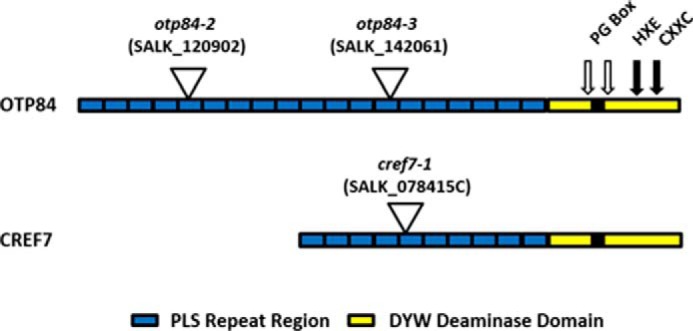
FIGURE 1.

PPR domain architecture for OTP84 and CREF7. Repeats in the PLS region are indicated by rectangles. Features in the DYW deaminase domain include the PG box, which is indicated as the solid black rectangle, and the open arrows indicate the position of truncations in OTP84. The positions of the zinc binding and catalytic glutamate residues (HXE, CXXC) are indicated with solid arrows. The location of T-DNA insertions is shown at the point of the triangle.
