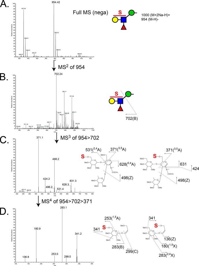
FIGURE 8.
MS spectra of the major sulfated, fucosylated O-mannose-linked glycan released from RPTPζ/phosphacan prepared from early postnatal mouse brains. A, enlarged view of full MS spectrum showing sulfated fucosylated O-mannosyl tetrasaccharide, m/z 954, from aqueous phase in the negative ion mode. B, MS2 spectrum of the precursor ion at m/z 954 in negative ion mode. The fragment ion at m/z 702 corresponds to B ion from the cleavage between GlcNAc and Man (sulfated fucosyl-LacNAc moiety). C, MS3 spectrum of the fragment ion at m/z 702 in negative ion mode. Ion peaks were assigned as the fragments of the indicated sulfated fucosyl-LacNAc structures. The most prevalent fragment ion at m/z 371 corresponds to cross-ring cleavage (3,5A) of a GlcNAc residue. D, further fragmentation of the ion at m/z 371 in MS3 produced fragment ions at m/z 253, 283, 299, and 341, which indicate sulfation of the Gal residue, and fragment ions at m/z 136, 180, 283, and 341, which indicate sulfation of the GlcNAc residue.

